
 精选
精选
在生物材料科学的浩瀚领域里,生物材料凭借其卓越的物理、化学和生物特性,已成为生物医学、制药和生物技术研究的焦点,具有不可替代的重要性。本期刊·见将为诸位介绍材料科学领域的高质量国际期刊Journal of Biomaterials Science, Polymer Edition。除了对期刊进行详尽的介绍外,还会向您介绍近一年高阅读量文章:

Print ISSN: 0920-5063
Online ISSN: 1568-5624
Journal of Biomaterials Science, Polymer Edition发表生物材料性能的基础研究,以及这些材料与生物体相互作用的机制,尤其关注分子和细胞层面的研究。
期刊涵盖的主题包括用于药物输送的聚合物、组织工程、以及生物体内大分子(如DNA、蛋白质等)。因此,Journal of Biomaterials Science, Polymer Edition融合了生物材料在生物医学、制药和生物技术领域的应用。
该期刊已被SCIE、 Ei Compendex 、MEDLINE等知名数据库收录。
影响因子
根据JCR显示,Journal of Biomaterials Science, Polymer Edition
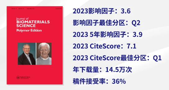
2023年影响因子为3.6
在工程:生物医学领域排名48/123
在材料科学:生物材料领域排名29/53
在高分子科学领域排名34/95
CiteScore
根据Scopus显示,Journal of Biomaterials Science, Polymer Edition
2023年CiteScore为7.1
在生物化学、遗传学和分子生物学:生物物理学领域排名30/152
在工程学:生物医学工程领域排名82/303
在材料科学:生物材料领域排名48/137
在化学工程:生物工程领域排名60/162
审稿周期
从提交稿件到获取初审意见,平均需要22天
获取首个同行评审决定,平均需要38天
稿件一旦接受后,在线出版平均需要12天
Journal of Biomaterials Science, Polymer Edition为混合开放获取期刊,支持作者以开放获取的模式发表文章,提升研究影响力。
编辑团队
Journal of Biomaterials Science, Polymer Edition的主编由俄亥俄州立大学的Stuart L. Cooper 教授、东京理科大学的Akihiko Kikuchi教授以及意大利特伦托大学的Antonella Motta教授共同担任,编委会由中国、日本、美国等地的行业翘楚组成。
主编介绍
Stuart L. Cooper

Stuart L. Cooper,俄亥俄州立大学杰出教授、美国国家工程院院士,主要从事聚合物物理、嵌段聚合物、离聚物、聚氨酯和生物材料领域的研究。
Akihiko Kikuchi

Akihiko Kikuchi,东京理科大学教授,研究方向为生物医学工程/生物材料科学与工程(生物材料、功能聚合物、药物输送系统、再生医学) 等领域。
Antonella Motta
Antonella Motta,意大利特伦托大学教授,研究方向为3D支架、生物相容性、生物材料、生物医学工程、生物聚合物、细胞培养等领域。
中国编委(部分介绍)
钟志远

钟志远,苏州大学特聘教授、药学院院长、国际创新药学院院长、放射医学与辐射防护国家重点实验室靶向放药中心主任。主要从事药物控制释放、肿瘤靶向治疗和免疫治疗研究。
计剑
计剑,科技部中国葡萄牙先进材料联合创新中心主任、浙江大学生物医用大分子研究所所长。主要从事生物医用材料的应用基础研究,在生命体系与材料界面的生物相容性和生物功能性方面开展了系统深入的研究。
作者分布
根据JCR显示,近三年在Journal of Biomaterials Science, Polymer Edition发文的国家中,发文量前三的国家/地区有:
中国
印度
伊朗
近三年,在Journal of Biomaterials Science, Polymer Edition发文的全球高校和科研机构中,发文较活跃的是:
印度贾米亚-哈姆达德大学
伊朗伊斯兰阿扎德大学
埃及知识库
均为开放获取文章,可免费查阅、下载
👇👇👇
近一年高阅读量文章
在明胶水凝胶支架上复制纳米级到微米级粗糙度的压印法:表面特征和对内皮化的影响
作者:Ali Salehi et al.
文章摘要:
Biologization of biomaterials with endothelial cells (ECs) is an important step in vascular tissue engineering, aiming at improving hemocompatibility and diminishing the thrombo-inflammatory response of implants. Since subcellular topography in the scale of nano to micrometers can influence cellular adhesion, proliferation, and differentiation, we here investigate the effect of surface roughness on the endothelialization of gelatin hydrogel scaffolds. Considering the micron and sub-micron features of the different native tissues underlying the endothelium in the body, we carried out a biomimetic approach to replicate the surface roughness of tissues and analyzed how this impacted the adhesion and proliferation of human umbilical endothelial cells (HUVECs). Using an imprinting technique, nano and micro-roughness ranging from Sa= 402 nm to Sa= 8 μm were replicated on the surface of gelatin hydrogels. Fluorescent imaging of HUVECs on consecutive days after seeding revealed that microscale topographies negatively affect cell spreading and proliferation. By contrast, nanoscale roughnesses of Sa= 402 and Sa= 538 nm promoted endothelialization as evidenced by the formation of confluent cell monolayers with prominent VE-cadherin surface expression. Collectively, we present an affordable and flexible imprinting method to replicate surface characteristics of tissues on hydrogels and demonstrate how nanoscale roughness positively supports their endothelialization.

作者:Sindi P. Ndlovu et al.
文章摘要:
Burn wounds are associated with infections, drug resistance, allergic reactions, odour, bleeding, excess exudates, and scars, requiring prolonged hospital stay. It is crucial to develop wound dressings that can effectively combat allergic reactions and drug resistance, inhibit infections, and absorb excess exudates to accelerate wound healing. To overcome the above-mentioned problems associated with burn wounds, SA/PVA/PLGA/Capparis sepiaria and SA/PVA/Capparis sepiaria nanofibers incorporated with Capparis sepiaria plant extract were prepared using an electrospinning technique. Fourier-transform infrared spectroscopy confirmed the successful incorporation of the extract into the nanofibers without any interaction between the extract and the polymers. The nanofibers displayed porous morphology and a rough surface suitable for cellular adhesion and proliferation. SA/PVA/PLGA/Capparis sepiaria and SA/PVA/Capparis sepiaria nanofibers demonstrated significant antibacterial effects against wound infection-associated bacterial strains: Pseudomonas aeruginosa, Enterococcus faecalis, Mycobaterium smegmatis, Escherichia coli, Enterobacter cloacae, Proteus vulgaris, and Staphylococcus aureus. Cytocompatibility studies using HaCaT cells revealed the non-toxicity of the nanofibers. SA/PVA/PLGA/Capparis sepiaria and SA/PVA/Capparis sepiaria nanofibers exhibited hemostatic properties, resulting from the synergistic effect of the plant extract and polymers. The in vitro scratch wound healing assay showed that the SA/PVA/Capparis sepiaria nanofiber wound-healing capability is more than the plant extract and a commercially available wound dressing. The wound-healing potential of SA/PVA/Capparis sepiaria nanofiber is attributed to the synergistic effect of the phytochemicals present in the extract, their porosity, and the ECM-mimicking structure of the nanofibers. The findings suggest that the electrospun nanofibers loaded with Capparis sepiaria extract are promising wound dressings that should be explored for burn wounds.
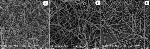 Figure 3. SEM images (a) NF7 (b) NF10 and (c) NF13.
作者:Doaa Elsayed Mahmoud & Nashiru Billa
文章摘要:
Biopharmaceutical and biomedical applications of chitosan has evolved exponentially in the past decade, owing to its unique physicochemical properties. However, further applications can be garnered from modified chitosan, specifically, depolymerized chitosan, with potentially useful applications in drug delivery or biomedicine. The use of microwave irradiation in depolymerization of chitosan appears to be more consequential than other methods, and results in modification of key physicochemical properties of chitosan, including molecular weight, viscosity and degree of deacetylation. In-depth review of such microwave-depolymerized chitosan and subsequent potential biopharmaceutical or biomedical applications has not been presented before. Herein, we present a detailed review of key physicochemical changes in chitosan following various depolymerization approaches, with focus on microwave irradiation and how these changes impact relevant biopharmaceutical or biomedical applications.
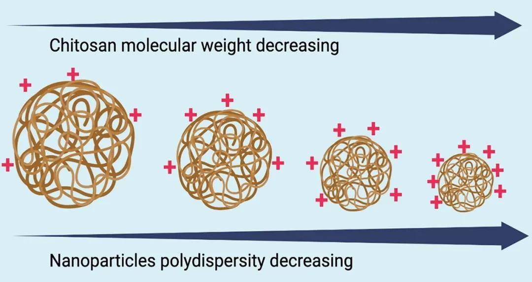
Figure 7. Effect of MW irradiation on chitosan molecular weight, nanoparticle size, polydispersity, surface morphology and charge.
"Jour - Know"
为帮助更多科研人员选择更加合适的期刊,Taylor & Francis推出专栏——刊·见,该专栏致力于为读者和广大科研人员带来Taylor & Francis旗下期刊的详细解读,从期刊的基本情况、编委阵容、社会影响力到审稿速度、高被引文章等实用信息,专栏将为您带来最详细的介绍,让您更加全面地了解Taylor & Francis旗下优秀的国际期刊,帮助更多中国卓越科研成果顺利在国际期刊上发表。
转载本文请联系原作者获取授权,同时请注明本文来自Taylor & Francis 学术服务科学网博客。
链接地址:https://wap.sciencenet.cn/blog-3574014-1474147.html?mobile=1
收藏