ВЉЮФ
дчЦкеяЖЯзЊвЦЕФЕквЛВН  ОЋбЁ
ОЋбЁ
||
дчЦкеяЖЯзЊвЦЕФЕквЛВН
жюЦН
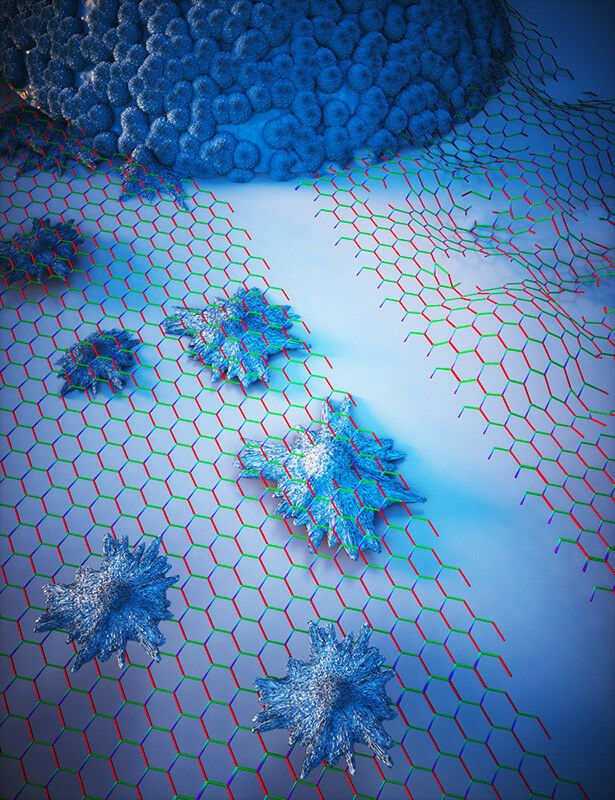
Stiff basement membranes promote metastasis Image: Ella Maru Studios.
ОнЕТЙњИЅРГБЄДѓбЇЃЈUniversity of FreiburgЃЉ2024Фъ3дТ4ШеЬсЙЉЕФЯћЯЂЃЌЕТЙњКЭЕЄТѓЕФбаОПаЁзщПЊЗЂСЫвЛжжаТЕФЗжЮіШЫРрЗЮВПЛљЕзФЄЃЈbasement membrane in human lungsЃЉЕФЗНЗЈЃЌЪЧТѕЯђдчЦкеяЖЯзЊвЦЕФЕквЛВНЃЈFirst Step Toward Early Diagnosis of MetastasisЃЉЁЃ
зЊвЦЪЧдьГЩЪЕЬхАЉЛМепЫРЭіТЪЕФжївЊдвђ;ШчЙћеяЖЯЮЊзЊвЦЃЌАЉжЂЛМепЕФдЄКѓЛсЯджјНЕЕЭЁЃЕНФПЧАЮЊжЙЃЌЛЙУЛгаПЩППЕФЗНЗЈРДдЄВтЮДРДзЊвЦЕФПЩФмадЁЃ
гЩИЅРГБЄДѓбЇвНбЇдКЃЈMedical Faculty, University of Freiburg, Freiburg, GermanyЃЉЕФГѕМЖНЬЪкРьГЖћЁЄТГЬиЃЈRaphael ReutenЃЉВЉЪПКЭЕТЙњФНФсКкгІгУПЦбЇДѓбЇ(Munich University of Applied Sciences, HM)ЕФКРПЫЁЄПЫРЭЩ-ЩмТќЃЈHauke Clausen-SchaumannЃЉНЬЪкСьЕМЕФРДздЕЄТѓКЭЕТЙњЕФПЦбЇМвЭХЖгЃЌЯждквбОТѕГіСЫдчЦкеяЖЯзЊвЦаЮГЩЕФЕквЛВН:ЫћУЧПЊЗЂСЫвЛжжгУЛЇгбКУЕФЗНЗЈРДЗжЮіШЫЬхЛљЕзФЄ-вђЮЊЫќЕФЛњаЕадФмЪЧзЊвЦЙ§ГЬжаЕФЙиМќвђЫиЁЃЯрЙибаОПНсЙћгк2024Фъ3дТ1ШевбОдкжјУћЕФПЦбЇдгжОЁЖздШЛавщЁЗЃЈNature ProtocolsЃЉЭјеОЗЂБэЁЊЁЊBastian Hartmann, Lutz Fleischhauer, Monica Nicolau, Thomas Hartvig Lindkær Jensen, Florin-Andrei Taran, Hauke Clausen-Schaumann, Raphael Reuten. Profiling native pulmonary basement membrane stiffness using atomic force microscopy. Nature Protocols, 2024. DOI: 10.1038/s41596-024-00955-7. Published: 01 March 2024. https://www.nature.com/articles/s41596-024-00955-7
ВЮгыДЫЯюбаОПЕФГ§СЫРДздИЅРГБЄДѓбЇКЭФНФсКкгІгУПЦбЇДѓбЇЕФбаОПШЫдБжЎЭтЃЌЛЙгаРДздЕТЙњФНФсКкЕФФЩУзПЦбЇжааФЃЈCenter for Nanoscience, Munich, GermanyЃЉЁЂЕЄТѓИчБОЙўИљДѓбЇЃЈUniversity of Copenhagen, Copenhagen, DenmarkЃЉЁЂЕЄТѓИчБОЙўИљЕФвНдКЃЈRigshospitalet, Copenhagen, DenmarkЃЉЕФбаОПШЫдБЁЃ
НЯШэЕФЛљЕзФЄИќФбПЫЗўЃЈSofter basement membranes are harder to overcomeЃЉ
РьГЖћЁЄТГЬиЫЕ:ЁАЮвУЧЯраХЃЌвдЯъЯИЗНАИЕФаЮЪНЗЂБэетжжЗНЗЈНЋЪЙЮвУЧИќНгНќгкзЊвЦаЮГЩЕФдчЦкеяЖЯЁЃЁБЪЙгУИУЗНАИЃЌЪРНчИїЕиЕФПЦбЇМвНЋФмЙЛШЗЖЈВЛЭЌЕФИіЬхЪЧЗёОпгаВЛЭЌЕФЛљЕзФЄСІбЇЃЌвдМАетаЉСІбЇЪЧЗёгызЊвЦаЮГЩгаЙиЁЃЛљЕзФЄЪЧЯИАћЭтЛљжЪ(ЯИАћЭтЕФЕААзжЪЭХ)ЕФвЛжжНсЙЙЃЌЫќАќЮЇзХЫљгаЕФбЊЙмЁЂаэЖрЦїЙйКЭжзСіЁЃ
Й§ШЅЃЌШЫУЧШЯЮЊетжжНсЙЙжЛЪЧАЉЯИАћБиаыНшжњздЩэЛњжЦПЫЗўЕФЦСеЯЁЃШЛЖјЃЌРьГЖћЁЄТГЬиКЭКРПЫЁЄПЫРЭЩ-ЩмТќдк2021ФъЕФвЛЦЊТлЮФжаЃЈin a paper from 2021ЃЉБэУїЃЌЛљЕзФЄЕФСІбЇЬиадБОЩэЪЧгАЯьАЉЯИАћзЊвЦЕФОіЖЈадвђЫиЃЌвђДЫвВЪЧгАЯьАЉжЂЛМепдЄКѓЕФОіЖЈадвђЫи:ЛљЕзФЄдНШэЃЌАЉЯИАћдНЩйЃЌДгЖјМѕЩйзЊвЦЃЌбгГЄЩњДцЦкЁЃ
АыздЖЏЛЏШэМўЗжЮіЃЈAnalysis by semi-automated softwareЃЉ
ПЦбЇМвУЧгыИЅРГБЄДѓбЇвНбЇжааФЕФИОВњПЦЃЈThe University Medical Center FreiburgЁЏs obstetrics and gynecology clinicЃЉКЭИчБОЙўИљвНдКЃЈRigshospitaletЃЉЕФВЁРэВПвЛЦ№ЃЌвдЯъЯИКЭЗНБугУЛЇЕФВНжшжИФЯЕФаЮЪНЗЂБэСЫШчКЮШЗЖЈШЫЬхЗЮЛљЕзФЄЕФЛњаЕЬиадЕФЗжЮіЙЄОпЁЃИУЭХЖгЛЙПЊЗЂСЫгУгкЗжЮіВтСПЪ§ОнКЭМјЖЈЛљЕзФЄЕФАыздЖЏШэМўЃЌЙЉПЦбЇНчЪЙгУЁЃЫцзХШЫЙЄжЧФмЕФЗЂеЙЃЌетИіШэМўдкЮДРДЛсБфЕУИќМгЧПДѓЁЃЩЯЪібаОПТлЮФЕФЕквЛзїепЁЂРДздФНФсКкгІгУПЦбЇДѓбЇ(HM)ЕФАЭЫЙЕйАВЁЄЙўЬиТќ(Bastian Hartmann)ЫЕ:ЁАвЛЕЉгаСЫИќШЋУцЕФЪ§ОнЃЌОЭПЩвдРћгУЛњЦїбЇЯАЪЕЯжЖдЛљЕзФЄЕФШЋздЖЏЪЖБ№ЁЃЁБ
ЛљЕзФЄЕФКёЖШжЛга100~400 nmЁЃЩњЮяЮяРэбЇМвКРПЫЁЄПЫРЭЩ-ЩмТќЫЕ:ЁАОЋШЗЖЈЮЛЫќдкзщжЏжаЕФЮЛжУЃЌВтСПЫќЕФЛњаЕадФмЃЌВЂДгжмЮЇзщжЏжаОЋШЗЕиЪЖБ№ЫќБОЩэОЭЪЧвЛИіЬиЪтЕФЬєеНЁЃЮвУЧФмЙЛЭЈЙ§ЙтбЇЯдЮЂОЕКЭдзгСІЯдЮЂОЕЕФНсКЯРДНтОіетИіЮЪЬтЁЃЁБаТЗНАИЯждкНЋетвЛГЬађРЉеЙЕНАќРЈШЫРрЗЮЛљЕзФЄЃЌЪЙЦфЖдУЛгаОбщЕФгУЛЇПЩааЁЃ
АЉжЂбаОПЕФЦфЫћЗЂЯжЃЈAdditional findings for cancer researchЃЉ
ПЦбЇМвУЧЦкД§гУЫћУЧЕФЗНЗЈЛёЕУАЉжЂбаОПЕФживЊЗЂЯжЁЃетЪЧвђЮЊОЁЙмжзСіжиЫмСЫЬхФкЕФаэЖрНсЙЙвдТњзуЦфашЧѓЃЌЕЋЖРСЂгкетаЉгыАЉжЂЯрЙиЕФБфЛЏжЎЭтЃЌЛљЕзФЄЕФИіЬхСІбЇЬиадЖдзЊвЦЙ§ГЬОпгажСЙиживЊЕФгАЯьЁЃвђДЫЃЌРьГЖћЁЄТГЬиЭЦВтЃЌЛљЕзФЄЕФФГаЉЛњжЦддђЩЯПЩФмЪЙФГаЉШЫИќШнвзЗЂЩњзЊвЦЁЃ
РьГЖћЁЄТГЬиВЉЪПзд2021ФъФъжавдРДвЛжБЕЃШЮИЅРГБЄДѓбЇЪЕбщгыСйДВвЉРэбЇКЭЖОРэбЇбаОПЫљЃЈThe University of FreiburgЁЏs Institute of Experimental and Clinical Pharmacology and ToxicologyЃЉЕФГѕМЖНЬЪкЃЌВЂСьЕМзХMateriTecture LabбаОПаЁзщЁЃЫћЕФбаОПжиЕуЪЧЯИАћЭтНсЙЙЖдЯИАћЙ§ГЬЕФгАЯьЃЌЬиБ№ЪЧжзСіЕФНјеЙЃЌФПЕФЪЧНЋЛљгкЛљжЪЕФеяЖЯКЭжЮСЦВпТдгІгУгкШеГЃСйДВгІгУЁЃ
КРПЫЁЄПЫРЭЩ-ЩмТќНЬЪкдкФНФсКкгІгУПЦбЇДѓбЇгІгУзщжЏЙЄГЬКЭдйЩњвНбЇжааФ(Munich University of Applied SciencesЁЏ Center for Applied Tissue Engineering and Regenerative Medicine)НјаабаОПЙЄзїЁЃЫћЕФзЈГЄЪЧдкФЩУзГпЖШЩЯЪЖБ№ЩњЮяВФСЯЕФЛњаЕЬиадЁЃАЭЫЙЕйАВЁЄЙўЬиТќЪЧКРПЫЁЄПЫРЭЩ-ЩмТќбаОПзщЕФВЉЪПЩњЃЌжївЊбаОПРћгУдзгСІЯдЮЂОЕЖдзщжЏНјааФЩУзСІбЇбаОПЁЃ
БОбаОПЕУЕНСЫЕТЙњАЭЗЅРћбЧжнПЦбЇКЭвеЪѕВПЭЈЙ§АЭЗЅРћбЧбаОПжааФ(CANTER)КЭАЭЗЅРћбЧбЇЪѕТлЬГ(BayWISS)НЁПЕбаОПВЉЪПСЊУЫ{ Bavarian State Ministry of Science and the Arts through the Bavarian Research Focus ЁЎHerstellung und biophysikalische Charakterisierung von dreidimensionalen Geweben (CANTER)ЁЏ and the Bavarian Academic Forum (BayWISS)ЁЊDoctoral Consortium ЁЎHealth ResearchЁЏ}ЬсЙЉЕФзЪН№ЁЃвВЕУЕНСЫРДздЕТЙњбаОПЛљН№ЛсЃЈGerman Research FoundationЃЉКЭЕЄТѓАЉжЂаЛс(Danish Cancer Society R204-A12454)ЕФзЪжњЁЃ
ЩЯЪіНщЩмЃЌНіЙЉВЮПМЁЃгћСЫНтИќЖраХЯЂЃЌОДЧызЂвтфЏРРдЮФЛђепЯрЙиБЈЕРЁЃ
Mammalian cells sense and react to the mechanics of their immediate microenvironment. Therefore, the characterization of the biomechanical properties of tissues with high spatial resolution provides valuable insights into a broad variety of developmental, homeostatic and pathological processes within living organisms. The biomechanical properties of the basement membrane (BM), an extracellular matrix (ECM) substructure measuring only ЁЋ100ЈC400 nm across, are, among other things, pivotal to tumor progression and metastasis formation. Although the precise assignment of the YoungЁЏs modulus E of such a thin ECM substructure especially in between two cell layers is still challenging, biomechanical data of the BM can provide information of eminent diagnostic potential. Here we present a detailed protocol to quantify the elastic modulus of the BM in murine and human lung tissue, which is one of the major organs prone to metastasis. This protocol describes a streamlined workflow to determine the YoungЁЏs modulus E of the BM between the endothelial and epithelial cell layers shaping the alveolar wall in lung tissues using atomic force microscopy (AFM). Our step-by-step protocol provides instructions for murine and human lung tissue extraction, inflation of these tissues with cryogenic cutting medium, freezing and cryosectioning of the tissue samples, and AFM force-map recording. In addition, it guides the reader through a semi-automatic data analysis procedure to identify the pulmonary BM and extract its YoungЁЏs modulus E using an in-house tailored user-friendly AFM data analysis software, the Center for Applied Tissue Engineering and Regenerative Medicine processing toolbox, which enables automatic loading of the recorded force maps, conversion of the force versus piezo-extension curves to force versus indentation curves, calculation of YoungЁЏs moduli and generation of YoungЁЏs modulus maps, where the pulmonary BM can be identified using a semi-automatic spatial filtering tool. The entire protocol takes 1ЈC2 d.
https://wap.sciencenet.cn/blog-212210-1424079.html
ЩЯвЛЦЊЃКДДаТЕФАЉжЂжЮСЦЪЙгУГЌЩљМЄЛювЉЮяАаЯђ
ЯТвЛЦЊЃКAIP:жаЙњдТЧђбљБОжаЕФЙшЭСНвЪОСЫдТЧђвўВиЕФРњЪЗ
ШЋВПзїепЕФОЋбЁВЉЮФ
ШЋВПзїепЕФЦфЫћзюаТВЉЮФ
ШЋВПОЋбЁВЉЮФЕМЖС
- • ЛљН№БОзгЁАЪнЩэЬсжЪЁБЃЌХфЭМВЛвЊВШет3жжПгЃЁ
- • зЃКиЃЁПЦбЇЭј2025ФъЖШЪЎМбВЉЮФЦРбЁЛюЖЏНсЙћНвЯў
- • ЮїББЙЄвЕДѓбЇеХЧягэ&еХКЭХє&ЭѕЬьЫЇЕШЃКИФадВЛПЩФцМќCOFsЪЕЯжИпаЇCO₂ЛЙд
- • ББОЉЪаПЦбЇММЪѕЮЏдБЛсЁЂжаЙиДхПЦММдАЧјЙмРэЮЏдБЛсЙњМЪКЯзїДІМАЙњМЪжааФвЛааЕНЗУMDPIББОЉЪаЭЈжнАьЙЋЪв
- • КЃбѓжаЕФАЕбѕ
- • ЩЯЮчЭЖЯТЮчUnder ReviewЃЌОоКУЭЖЕФЫФЧјSCIРДИхОЭЪеЃЌ0АцУцЗбЃЁ